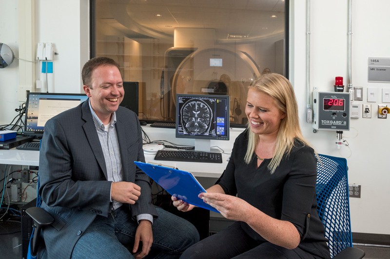
UD researchers study biological roots for adolescent risk-taking
As any parent will tell you, no two children behave in exactly the same way. It is part of what makes each individual unique.
So, why do some adolescents take more risks than others?
University of Delaware Biomedical Engineer Curtis Johnson and graduate student Grace McIlvain think they may have an idea.
The part of the brain that makes adolescents want to take risks is called the socioemotional system. The brain’s cognitive control center, meanwhile, is what helps prevent adolescents from acting on these impulses.
In a recently published paper in NeuroImage, Johnson and McIlvain suggest that these two centers in the brain physically mature at different rates and that adolescents with large differences in the rate of development between these two brain regions are more likely to be risk-takers. Further, the research team theorizes that it is the brain’s fundamental structure that drives these risk-taking and control tendencies.
What makes this study unique is that the UD researchers and their collaborators used a technique called magnetic resonance elastography (MRE) to safely measure the mechanical properties of the brain tissue as a measure of brain development, rather than activation of those two regions.
Elastography is a method of imaging mechanical properties of tissues using a magnetic resonance imaging (MRI) scanner. Simply put, the researchers take snapshots of how the brain deforms — or bends — as it is vibrated under low frequencies, and then put those images through a specific algorithm to reverse engineer what is happening. Johnson explained that MRE vibration is safe for all ages and provides less movement than naturally occurs in the brain. It also offers less vibration than other devices designed for children, such as vibrating rockers.
Johnson likened the process to any other material testing and said the research team’s knowledge of how tissue deforms helps them interpret what is happening under different vibrations. In adults, MRE techniques have become popular for studying diseases, such as Alzheimer’s, with research showing relationships between memory and cognitive performance.
“MRE techniques do not replace other aspects of studying brain development, but they may provide a more sensitive, objective way to look at the brain’s wiring,” said Johnson, an assistant professor in the Department of Biomedical Engineering.
Mapping adolescent brain development
This is not the first time that researchers have looked at how two brain regions interact to form a certain output. But most of this work has been done using functional MRI (fMRI), where study participants are placed in the scanner and given a real-time task, and the researchers watch which areas of the brain light up to determine what areas of the brain relate to that task.
Johnson’s research group was an early pioneer in using MRE techniques to make high-resolution three-dimensional maps that enable scientists to look at specific regions of the brain. The intensity of every 3D pixel in an image has meaning. For example, bright colors indicate high stiffness, which, in this case, indicates a measure of developmental maturity.
Looking at these features of the brain in their work, the researchers found that it wasn’t the socioemotional or the cognitive control center alone, but the combination of the two centers of the brain working together at a specific age or point in time that was the definitive factor in risk taking.
“So, there is this period during adolescence where the part of the brain that makes you want to take risks is more mature than the part of the brain that suppresses those impulses,” said McIlvain, who began working on the project as an undergraduate summer researcher in 2016 and is now a third-year doctoral student in biomedical engineering.
“If we can identify individuals who are more likely to take risks, based on the biological composition of their brain, or maybe groups of individuals, it might inform strategies for prevention.”
Prior to this project, little MRE research had measured brain stiffness in children. Earlier work in 2018 by McIlvain showed the outside of the brain appears softer in adolescents than adults, whereas the inside of the brain appears stiffer in adolescents than adults. According to Johnson, this aligns with the known developmental trajectory where the inside of the brain develops first and the outside, the cortex, develops later.
The work grew out of Johnson’s previous collaborative research with Eva Telzer, a psychology professor at University of North Carolina and co-author of the paper, and leverages the advanced MRI capabilities at UD’s Center for Biomedical and Brain Imaging. Today, researchers in the Johnson lab develop all aspects of this MRE technique, from how to safely vibrate the head in the scanner to how to write the software to acquire the data to methods for turning the data into images that are translated into mechanical properties.
While the research team’s previous work has shown differences in the brain function of typically developing children and those with conditions, such as cerebral palsy, this is the first time the researchers have shown a relationship with function in healthy children. But there are still more questions than answers.
For example, Johnson said currently there are no good measures for saying when the brain is mature or even for how to define brain health. And while the research team has made connections between how stiffness of the adolescent brain’s socioemotional system and cognitive control center interrelate and support risk taking, there are other things they don’t know, like how these regions of the brain are affected by things like socioeconomic status, early life trauma or early education.
A big focus of the work is making the MRE scan faster. The scan currently takes over six minutes, which can be difficult for children with disabilities or those who are very young.
“We’d like to complete the scan in under a minute — less time than half a song from a Disney movie — before a child loses interest and thinks about moving,” said Johnson.
Next steps in the research include scanning kids as young as age 5, including those with autism. The hope is to create a robust data set to explore how brain mechanical properties change from age 5 to age 30, generally considered to be the end of adolescence. Among other things, they hope to use this data to better understand how children with disabilities fit into that developmental curve.
“Right now, there is no standard way to diagnose autism, no targeted treatment plan or metrics for measuring whether intervention is helping,” said McIlvain, who recently was awarded an National Institutes of Health fellowship to study brain stiffness in children with autism. “If we can understand how the mechanical properties of the brain are affected in someone with autism, we can start to answer some of those questions.”
| Photo by Kathy F. Atkinson

