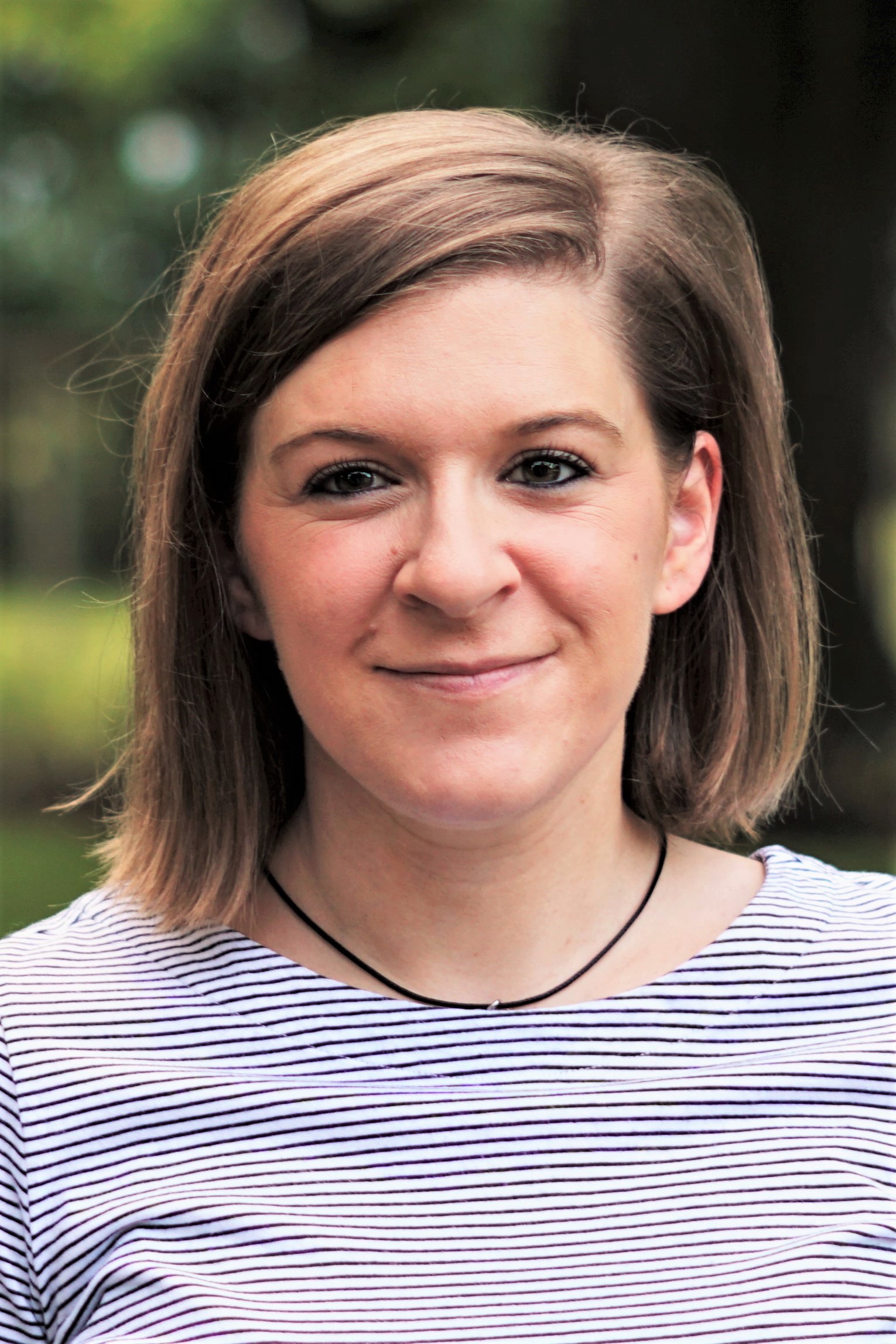
BME PhD Candidate, Keely A. Keller, will be defending her dissertation
TITLE:
Techniques for Enhanced Biomimicry of In Vitro Vascular Models
When: Friday, July 24, 2020 @ 1:00 pm
Where: Zoom Meeting ID: 975 9790 7482
Password: Keller
Committee Chair: John Slater, PhD
Committee: Jason Gleghorn, PhD., Kelvin Lee, PhD., Christopher Martens, PhD.
ABSTRACT:
With the potential to lie dormant for years after extravasating into a secondary organ, circulating tumor cells (CTCs) that shed into the bloodstream from a primary tumor during metastasis represent a significant long-term health concern for cancer patients. While the organ-specific affinity of CTCs, termed organotropism, is not yet fully understood, a better insight into CTC behavior within the microvasculature of different organs could provide potential pharmaceutical targets for prevention of metastatic relapse. In vitro, human-derived vascular models have enormous potential in elucidating CTC adhesion and entrapment within vasculature, and interactions with, and disruption of endothelial cell monolayers, due to their enhanced imaging capabilities, and environmental control compared to animal models. Therefore, the overarching goal of this research was to develop methods and tools for the creation of organ-specific in vitro vascular models with regards to recapitulating microvascular architecture, fluid flow profiles, and barrier function.
The first objective of this research was to recapitulate the complex organ-specific architecture of microvasculature in in vitro constructs, as vascular branch points, narrowings, and overall network tortuosity can influence CTC entrapment. To do so, we developed image-guided, laser-induced hydrogel degradation (LIHD) for the creation of highly resolved microfluidic channels within both natural (collagen) and synthetic (PEGDA) hydrogels that support endothelial cell growth. Using this technique, we demonstrate the exact replication of mouse brain vascular architecture, and create a construct with two independent and intertwining microfluidic channels to recapitulate the close interaction between cardio and lymphatic vasculature. We also demonstrate how LIHD can be used to locally control the concentration and porosity of hydrogels, and use this for size-based separation of several biomolecules. These results highlight the wide range of applications and design freedom afforded by LIHD.
The second objective of this research was to ensure physiologically relevant fluid flow profiles in complex in vitro vascular models. The architecture of a microvascular network has an influence on the fluid flow, with vascular branching, bifurcations, and curvature generating gradients and localized disturbances in wall shear stress which can then locally influence endothelial cell behavior. Using pressure-head and syringe pump driven fluid flow through LIHD-generated channels and networks in microfluidic device-housed hydrogels, we demonstrate several techniques for visualizing interstitial and luminal fluid flow. To recapitulate in vivo fluid flow within in vitro vascular models, we used particle image velocimetry to validate custom microfluidic network augmentation software within LIHD-generated devices. We also demonstrate the ability to recapitulate bulk properties of a capillary bed using synthetic biomimetic microfluidic networks. Finally, we present preliminary efforts examining the entrapment of model CTCs in LIHD-generated channels.
The final objective of this research was to improve the vascular barrier function of in vitro vascular models to enable accurate study of CTC extravasation and breaching of these barriers in the future. We postulated that organ-specific endothelial cells would form more robust cell-cell junctions if cultured on an organ-specific basement membrane-mimetic formulation of cell-adhesive peptides, as opposed to standard culture on the ubiquitous RGDS cell-adhesive peptide. We show that human lung-derived microvascular endothelial cells, when cultured in the lung BM mimetic peptide formulation, form vessels that are less permeable to 10 kDa dextran, and that appear to generate more intercellular force. The results presented will guide the smart design of organ-specific, basement membrane-mimetic cell culture substrates within in vitro vascular models for enhanced vascular barrier function.
Overall, this dissertation advances the state-of-the-art in generating organ-specific in vitro vascular devices by recapitulating organ-specific microvascular architecture, fluid flow profiles, and barrier function. These tools and techniques will be critical in determining how and why CTCs colonize particular organs over others, and will aid in the discovery of anti-metastatic pharmaceuticals.

